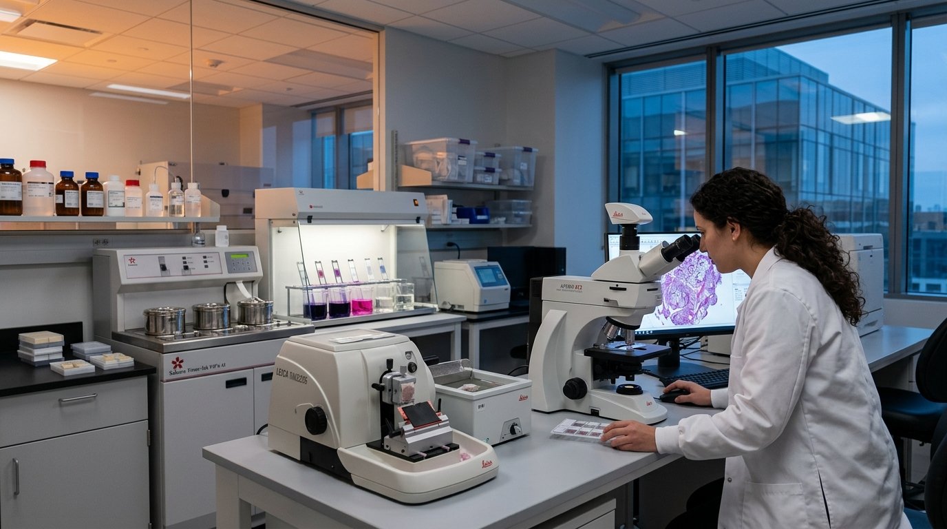
End-to-end tissue processing, embedding, precision microtomy, differential histological staining, and high-resolution digital photomicrography — providing cellular-level evidence of drug-induced effects, disease pathology, and treatment response in preclinical study tissues.
Histopathological evaluation provides irreplaceable microscopic evidence of tissue-level changes induced by disease, therapeutic intervention, or toxic insult. Our histopathology division offers a complete processing pipeline from fresh tissue fixation to stained slide preparation and semi-quantitative scoring — making it an essential complement to biochemical and pharmacological endpoints in preclinical studies.
Leica Rotary MicrotomeFreshly harvested organs (liver, kidney, pancreas, lung, brain, spleen, heart, intestine) fixed in 10% Neutral Buffered Formalin (NBF) for 24–48 hours. Processed through graded ethanol dehydration series (70%, 80%, 90%, 95%, 100%), xylene clearing, and paraffin infiltration using an automated tissue processor. Paraffin blocks prepared using embedding station with stainless steel molds.
Paraffin blocks sectioned using calibrated Leica rotary microtome at 4–5 µm thickness (adjustable 1–50 µm). Sections floated on warm water bath (37–40°C) and mounted on silane-coated glass slides. Dried overnight in hot air oven at 58°C for optimal adhesion prior to deparaffinization and staining.
Routine H&E staining for general tissue morphology assessment. Hematoxylin stains nuclei basophilic (blue-purple); eosin stains cytoplasm and extracellular matrix acidophilic (pink). Suitable for evaluation of cellular architecture, inflammatory infiltration, necrosis, hepatocyte vacuolation, glomerular changes, islet morphology, and general organ pathology.
Masson's Trichrome (MT): Collagen fiber quantification and fibrosis assessment (liver, kidney, cardiac). Collagen = blue, muscle = red, nuclei = dark brown.
Congo Red: Amyloid deposit detection with apple-green birefringence under polarized light.
Periodic Acid-Schiff (PAS): Glycogen and polysaccharide detection in hepatic and renal tubular cells.
von Kossa: Calcium phosphate deposit identification in renal urolithiasis studies.
Oil Red O: Lipid droplet detection in hepatosteatosis (fresh cryosections required).
High-resolution digital images captured using trinocular compound microscope with calibrated camera at 4×, 10×, 40×, 100× (oil immersion) objectives. Images processed with calibration bar using image analysis software. Semi-quantitative histopathological scoring (0 = normal, 1 = mild, 2 = moderate, 3 = severe) by trained assessor. Representative photomicrographs selected for each experimental group.
Organs harvested at sacrifice, weighed, and immediately immersed in 10% NBF. Representative portions trimmed for processing.
Fixed tissues placed in labeled cassettes and processed through dehydration, clearing, and paraffin infiltration cycle overnight (12–18 hours).
Processed tissues oriented and embedded in paraffin using embedding station. Blocks trimmed and labeled with study ID.
4–5 µm serial sections cut by calibrated microtome, floated, mounted on glass slides, and dried in oven.
Slides deparaffinized, rehydrated, and stained per protocol (H&E, MT, special stains). Coverslipped with DPX mountant.
Stained slides examined, representative images captured at multiple magnifications, and scoring table compiled with interpretive notes.
Discuss your histopathology requirements
Share your tissue details and we'll discuss the best staining approach.