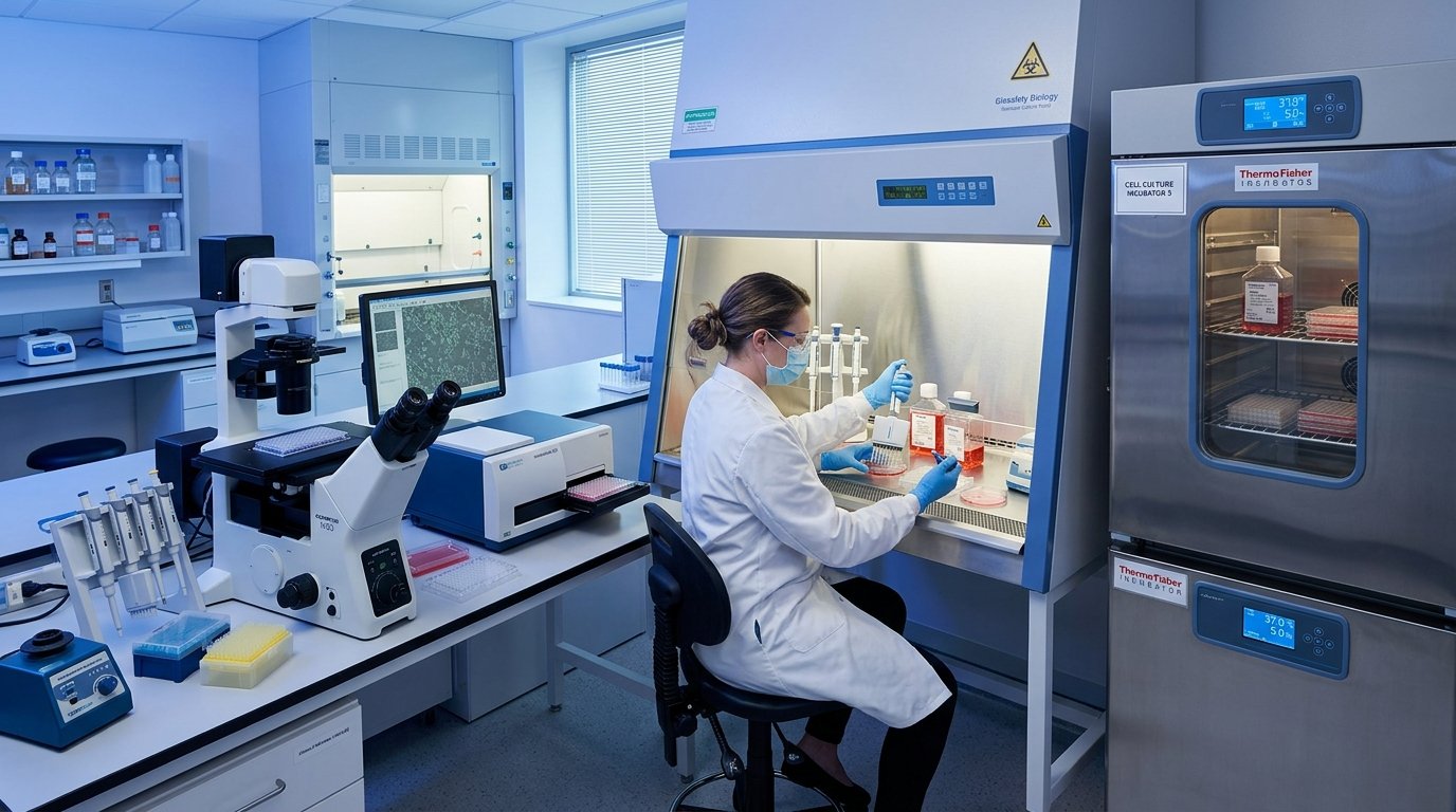
Standardized mammalian cell culture maintenance and high-throughput in-vitro cytotoxicity, apoptosis, oxidative stress, and molecular biology assays — providing mechanistic insights into test substance activity at the cellular level.
In-vitro cell-based assays provide the primary mechanistic evidence for antiproliferative, cytotoxic, apoptotic, and cytoprotective activities of test compounds. Our cell biology laboratory maintains authenticated human cancer and normal cell lines in dedicated CO₂ incubators, using rigorously validated assay protocols that generate reproducible, quantitative data suitable for publication in peer-reviewed journals.
Class II BSC Cell Culture SuiteCertified human cancer cell lines (MCF-7 breast, HeLa cervical, A549 lung, HT-29 colon, HepG2 liver) and non-cancerous control cell lines (NIH-3T3, L929). Maintained in DMEM/RPMI-1640 with 10% FBS, penicillin-streptomycin, in 37°C/5% CO₂ incubators. Mycoplasma-free status confirmed. Trypan blue exclusion viability assessment and passage documentation maintained.
Colorimetric assessment of mitochondrial NAD(P)H-dependent cellular oxidoreductase activity using 3-(4,5-dimethylthiazol-2-yl)-2,5-diphenyltetrazolium bromide (MTT). Dose-response curves generated across 6–8 concentrations (serial 2-fold dilution), with non-linear regression IC50 calculation using GraphPad Prism. Selectivity Index (SI = IC50 normal / IC50 cancer) calculated where applicable. Standard: Doxorubicin, Cisplatin.
Detection of early and late apoptotic cell populations using Annexin V-FITC and Propidium Iodide (PI) double staining followed by fluorescence microscopic analysis. Quantitative cell population categorization: live (Annexin⁻/PI⁻), early apoptotic (Annexin⁺/PI⁻), late apoptotic (Annexin⁺/PI⁺), and necrotic (Annexin⁻/PI⁺). Representative fluorescence photomicrographs provided.
Measurement of intracellular reactive oxygen species (ROS) accumulation using 2',7'-dichlorodihydrofluorescein diacetate (DCFH-DA) fluorescent probe. Cells loaded with DCFH-DA are treated with test compound and H₂O₂ (positive control). Relative fluorescence units (RFU) quantified by fluorescence microplate reader at excitation/emission 485/535 nm. Results expressed as % ROS generation vs. untreated control.
Acridine Orange (AO) and Propidium Iodide (PI) dual fluorescence staining for visual differentiation of viable, apoptotic, and necrotic cell populations under inverted fluorescence microscope. AO intercalates into double-stranded DNA (live cells fluoresce green); PI penetrates membrane-compromised cells (late apoptotic/necrotic cells fluoresce red). Documented with representative fluorescence photomicrographs.
Caspase-3/7 apoptotic pathway activation measured by colorimetric caspase substrate cleavage assay (Ac-DEVD-pNA). Cell cycle distribution (G0/G1, S, G2/M phase arrest) assessed by PI staining and DNA content analysis via fluorescence microscopy or microplate reader. Cell cycle data interpreted relative to compound mechanism and dose.
Target cell lines seeded in 96-well plates at defined cell density. 24h pre-incubation for monolayer attachment at 37°C/5% CO₂.
Serial dilutions of test substance prepared in DMSO:culture medium (DMSO ≤ 0.1%). Treated cells incubated for 24h/48h/72h per protocol.
MTT/DCFH-DA/Annexin V reagent added per assay protocol. Formazan crystals solubilized in DMSO for MTT; fluorescence read directly for ROS.
Multimode microplate reader records OD (570nm for MTT) or fluorescence emission. Minimum 3 independent replicates per concentration.
Non-linear regression (log concentration vs. % viability) in GraphPad Prism. One-way ANOVA + Dunnett's test. p < 0.05 threshold.
Discuss your cell biology assay requirements
Connect with our cell biology team to design your assay panel.