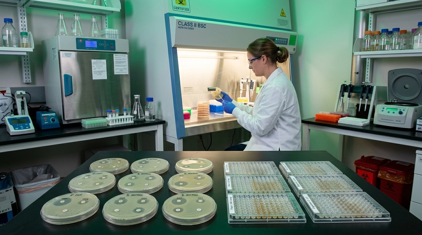
Standardized in-vitro antimicrobial susceptibility evaluation including Minimum Inhibitory Concentration (MIC), Minimum Bactericidal Concentration (MBC), disk diffusion zone of inhibition, biofilm inhibition, and time-kill kinetics — conducted against clinically relevant bacterial and fungal pathogens per CLSI guidelines.
Antimicrobial research is a critical evaluation for any molecule intended to address bacterial or fungal infections — whether from natural product chemistry, synthetic drug development, or novel formulation research. Our microbiology laboratory performs well-characterized susceptibility testing against ATCC reference strains and clinically relevant isolates, using CLSI-standardized protocols that are universally accepted in scientific publications and regulatory submissions.
Class II BSC Microbiology SuiteMueller-Hinton agar plates inoculated with standardized bacterial suspension (0.5 McFarland turbidity = ~1.5 × 10⁸ CFU/mL). Test compound-impregnated filter disks (6 mm diameter) placed on agar surface. Plates incubated at 37°C for 18–24 hours. Zone of inhibition (ZOI) measured in millimeters. Results compared against positive control (antibiotic standards) and negative controls (DMSO/solvent vehicle). Breakpoint interpretation per CLSI M100.
Minimum Inhibitory Concentration (MIC) determined using 96-well broth microdilution format as per CLSI M07 guidelines. Two-fold serial dilutions of test compound prepared in Mueller-Hinton broth (bacteria) or RPMI-1640/Sabouraud broth (fungi). Inoculated with 5 × 10⁵ CFU/mL, incubated 18–24 hours (bacteria) or 48–72 hours (fungi). MIC defined as lowest concentration with no visible growth. Resazurin indicator (blue-to-pink color change) used as viability indicator for microplate reading.
Sub-culturing of wells showing no visible growth from MIC assay onto drug-free Mueller-Hinton agar plates. After 18–24h incubation, colony counts performed. MBC defined as lowest concentration killing ≥ 99.9% of original inoculum. MBC/MIC ratio calculated to classify compound as bactericidal (ratio ≤ 4) or bacteriostatic (ratio > 4). Essential for antibiofilm and drug mechanism interpretation.
Biofilm formation quantified using crystal violet (CV) staining in 96-well polystyrene plates. Bacteria inoculated and allowed to form biofilm (24–48h at 37°C without agitation). Planktonic cells removed by washing; adherent biofilm stained with 0.1% CV. Biofilm biomass solubilized with 95% ethanol; absorbance read at 570 nm. Test compound effect expressed as % biofilm inhibition relative to untreated control.
Evaluation of potential synergistic, additive, or antagonistic interactions between test compound and standard antibiotics (Ampicillin, Ciprofloxacin, Fluconazole) using checkerboard microdilution assay. Fractional Inhibitory Concentration Index (FICI) calculated: synergy (FICI ≤ 0.5), addition (0.5 < FICI ≤ 1.0), indifference (1 < FICI ≤ 2), antagonism (FICI > 2). Used to support combination antibiotic formulation research.
ATCC reference strains revived and grown on appropriate media. Working stocks prepared and McFarland turbidity standardized.
Stock solution prepared in DMSO; serial two-fold dilutions prepared in sterile broth. Solvent control included at highest DMSO concentration used.
Standardized bacterial suspension added to dilution series. Plates sealed and incubated at 37°C for 18–24 hours.
Visual or resazurin-aided reading of growth inhibition. MIC recorded; sub-culturing performed for MBC determination.
MIC/MBC values tabulated. Disk diffusion ZOI measurements recorded. Comparison with standard antibiotics. Complete testing report generated.
Specify organisms and assay type required
Our microbiology team is available to discuss your testing panel requirements.